
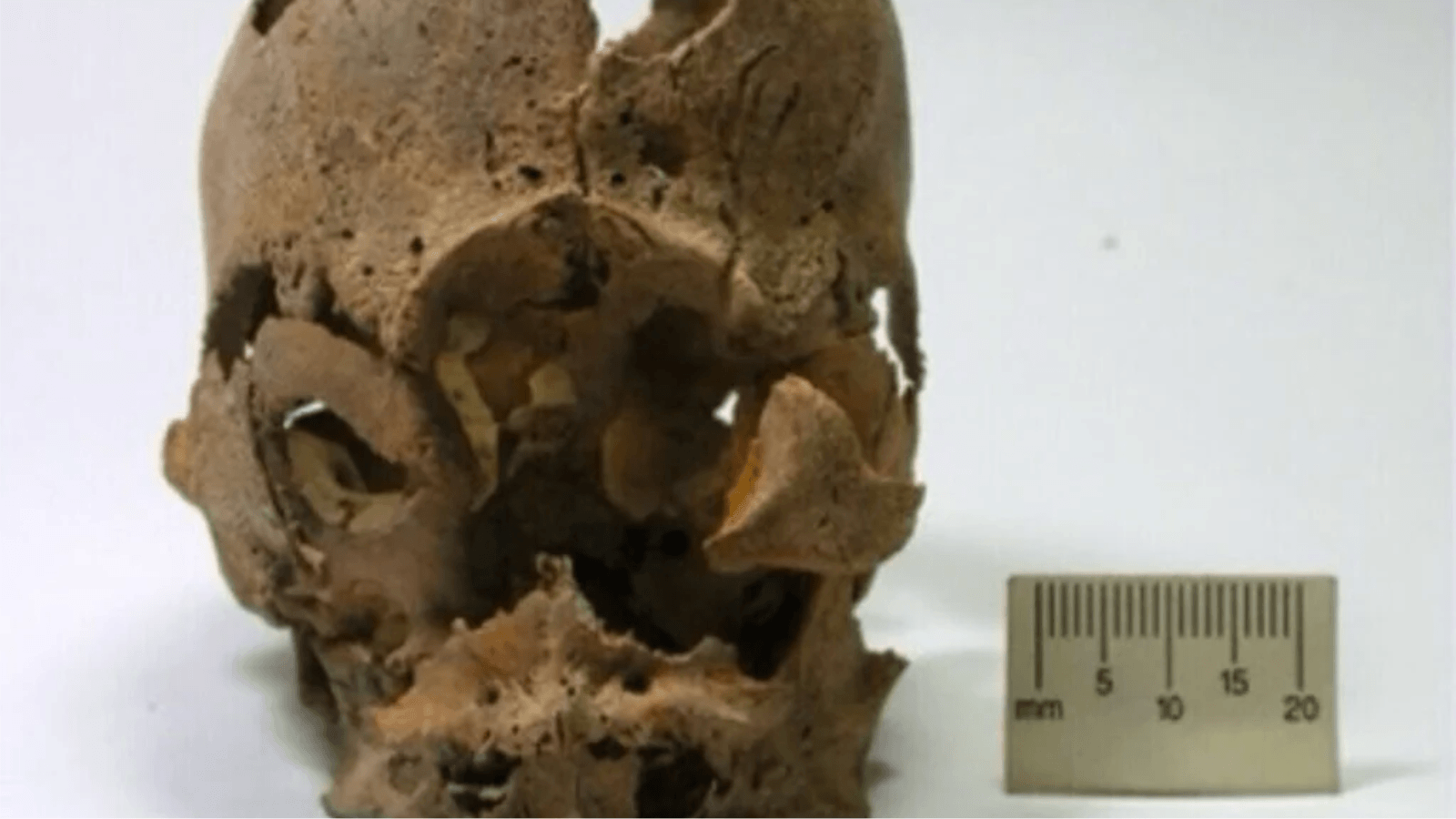
A 1,600-year-old infant buried in southern Peru had a single eye socket in the center of her skull, a severe congenital anomaly whose genetic cause has now been identified through ancient DNA analysis. The child carried an extra copy of chromosome 13, a condition associated with major abnormalities in brain and facial development. Her remains were found alongside another infant in a ceramic vessel, raising additional questions about burial practices in the ancient Nasca culture.
The findings, published in the Proceedings of the National Academy of Sciences (PNAS), provide genetic evidence for a rare condition known as cyclopia. They also show that the infant was buried according to a funerary tradition commonly practiced for children in the region, despite her unusual physical characteristics.
Two Infants Found in a Nasca Burial
The remains came from Jauranga, an archaeological site in the Palpa Valley of southern Peru. Originally occupied during the earlier Paracas period, the site later became a burial ground for communities associated with the Nasca culture.
Archaeologists documented 31 graves dating to approximately AD 325–440. One of them, designated Burial 25, contained the articulated skeletons of two infants placed one above the other inside a ceramic vessel. No additional grave goods were recorded.
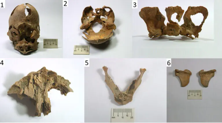
Both infants were genetically identified as female, but their skeletal remains differed considerably. One child, designated JA13A, had an unusually small skull with a single central eye socket instead of two separate orbital cavities. Her nasal bones were absent, and several structures forming the middle of the face had failed to develop normally.
The second infant, JA13B, showed no comparable skeletal abnormalities.
Ancient DNA Identifies Trisomy 13
The research team, led by Elsa Tomasto-Cagigao of the Pontifical Catholic University of Peru and Lars Fehren-Schmitz of the University of California, Santa Cruz, combined skeletal examinations with ancient DNA sequencing to investigate the condition.
JA13A’s craniofacial anatomy was consistent with cyclopia, the most severe form of a developmental disorder called alobar holoprosencephaly. The condition occurs when the embryonic forebrain fails to separate properly into two hemispheres, disrupting the development of the eyes and central facial structures.
Cyclopia affects approximately one in 100,000 births and is generally fatal before or shortly after delivery.
Genetic analysis provided an explanation for the infant’s unusual anatomy. Researchers detected three copies of chromosome 13 rather than the usual two, identifying a chromosomal disorder known as trisomy 13, or Patau syndrome.
To verify the finding, the team used three independent analytical approaches and compared the infant’s genetic data with those of 250 ancient South American individuals. All three methods supported the presence of an additional chromosome 13, establishing a strong genetic link between the disorder and the skeletal abnormalities.
The examination also identified differences beyond the skull, including unusually curved shoulder blades and two malformed vertebrae.
Did the Infant Survive After Birth?
The child’s skeletal development presented another question. Dental formation suggested a developmental age equivalent to approximately two to four months after birth, while measurements of the long bones corresponded more closely to a fetus at 36 to 40 weeks of gestation.
This discrepancy raises the possibility that the infant survived beyond the immediate period surrounding birth. However, the researchers emphasize that severe developmental conditions such as trisomy 13 can disrupt normal bone and tooth growth, making conventional age estimates unreliable.
Postnatal survival therefore remains plausible but unconfirmed. The evidence cannot establish how long the infant lived or whether she received care after birth.
Ancient DNA also showed that the two children belonged to different maternal lineages. Although they shared the same burial vessel, researchers could not establish why they were buried together or whether another relationship connected them.
A Familiar Burial Tradition Despite an Unusual Condition
The archaeological context provides another important aspect of the discovery. Ceramic vessel burials accounted for approximately one-quarter of documented Nasca graves in the Palpa region and were predominantly used for children younger than six years old.
Archaeologists have documented similar uses of pottery vessels in other ancient societies, including a Roman infant burial discovered in Britain alongside the remains of two birds. Although the discoveries belong to different cultural traditions, both illustrate the use of ceramic containers in childhood burials.
At Jauranga, Burial 25 followed the established Nasca funerary tradition. Its simplicity and absence of grave offerings were consistent with other known childhood burials, suggesting that JA13A’s physical differences did not result in archaeologically distinguishable funerary treatment.
This does not mean that the community necessarily understood or accepted her condition in a particular way. Burial customs cannot establish how an individual was perceived, treated, or cared for during life.
Archaeological evidence from other Andean societies demonstrates that responses to physical differences varied across cultures and periods. Moche artistic representations depict individuals with congenital differences, while skeletal evidence elsewhere indicates that some people with substantial physical impairments received assistance.
However, the researchers caution against applying those examples directly to Nasca society.
The Jauranga discovery demonstrates how genetic analysis can identify the biological origins of unusual skeletal conditions while archaeology provides evidence of their social and funerary context. In this case, the genetic diagnosis is clear, but the circumstances of the infant’s life and her shared burial remain unresolved.
Cover Image: The 1,600-year-old skull of an infant from Peru’s Nasca culture, showing a single central eye socket associated with cyclopia and trisomy 13. Credit: Tomasto-Cagigao et al., PNAS (2026).