
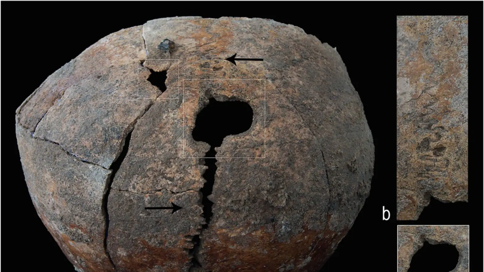
A circular opening in the skull of an adolescent buried in Bronze Age Myanmar may preserve evidence of a remarkable medical intervention. Its healed edges suggest that the young person survived the procedure for months—and possibly years—while living with a damaged jaw and a serious bone disorder.
The discovery is not simply a story of ancient surgery. Despite injuries that would have made eating and daily life difficult, the adolescent reached approximately 14 and a half years of age without clear signs of impaired growth or prolonged nutritional stress. Researchers believe such survival would probably have required sustained care from other members of the community.
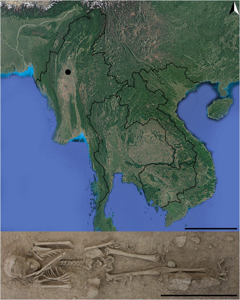
The findings come from a new osteobiographical study of Burial 3b at Nyaung’gan, a prehistoric cemetery in north-central Myanmar. Published in the International Journal of Osteoarchaeology, the research reconstructs the adolescent’s life from an unusually complex combination of trauma and disease.
A burial filled with signs of belonging
The adolescent lived sometime between 1000 and 700 BCE, during Myanmar’s early-to-middle Bronze Age, and was buried inside a hollowed-out log coffin. The grave contained 32 small pottery vessels, more than 100 semiprecious stone beads and a bone bracelet.
Although the offerings broadly resembled those placed in other graves at Nyaung’gan, the adolescent’s beads were unusually varied in material, size and form. Directly above the body were the primary burial of a dog and the secondary burial of a young adult man’s skull. Chemical evidence indicates that the man had grown up outside the area.
Isotope values recorded in Burial 3b, by contrast, resemble those of other people from Nyaung’gan. The results suggest that the adolescent grew up locally and consumed a similar diet, placing them firmly within the community rather than identifying them as an outsider.
Researchers Anna Willis, Baptiste Pradier, Thu Thu Win and T. O. Pryce examined the skeleton macroscopically and through radiographs taken at a private hospital in Mandalay. Because access to advanced imaging was limited when the remains were studied in 2016, their conclusions rest on the combined pattern of visible and radiographic changes rather than a single definitive test.
Two openings with different histories
The skull contained two major lesions. One was an irregular depression accompanied by radiating fractures, interpreted as evidence of a severe blow sustained during childhood. Part of the damaged bone appears to have died and gradually been absorbed as the injury healed.
The second opening was markedly different. It was circular, crossed the skull’s midline and had smooth, remodelled margins. Its shape and healing pattern made infection, tumours and other natural biological processes less convincing explanations.
The researchers concluded that these features are more consistent with intentional trepanation, a procedure in which part of the skull is deliberately opened or removed. Bone remodelling around the opening indicates that it was not produced at or after death. If the interpretation is correct, the adolescent survived the intervention for at least several months and potentially for years. Evidence of similarly risky cranial intervention has also been identified in the 4,000-year-old skull of a child buried at Djarkutan in Uzbekistan.
Why the procedure was undertaken cannot be determined. It may have been intended to relieve swelling or pressure associated with the earlier head injury and a resulting haematoma. Another possibility concerns the partial premature fusion of the adolescent’s cranial sutures.
Whether the person who performed the operation understood pressure inside the skull in anything resembling modern clinical terms remains unknowable. The healed opening nevertheless points to deliberate intervention, considerable practical skill and some form of postoperative care.
A jaw forced to adapt
The earlier trauma may also have affected the adolescent’s jaw. Uneven muscle attachments, altered tooth wear, reactive bone and changes to a developing molar root indicate that chewing had been abnormal for years.
The researchers consider a superolateral dislocation of the right temporomandibular joint—the joint connecting the lower jaw to the skull—the most probable explanation. Fractures of the jaw and its coronoid process may have occurred during the same traumatic event. A later case from Siberia offers a striking comparison, where researchers identified a 2,500-year-old attempt to reconstruct a damaged temporomandibular joint.
The skeletal changes began before the adolescent was approximately 10 and a half to 11 and a half years old, early enough to disrupt the formation of an adult tooth root. The chewing muscles subsequently adapted in different ways on either side of the face, suggesting long-term compensation for a badly misaligned jaw.
Red staining on the front teeth provides another intriguing but uncertain detail. The study raises the possibility that the adolescent was given areca, a plant traditionally used in the region for its stimulant and pain-relieving properties. Areca can stain teeth red, although the discoloration cannot by itself demonstrate medicinal use.
Disease recorded throughout the skeleton
Changes in the arms and legs reveal an additional health problem. The long bones were slightly curved, their ends were widened and their density was reduced. Poorly mineralized new bone and possible insufficiency fractures are consistent with osteomalacia related to vitamin D deficiency.
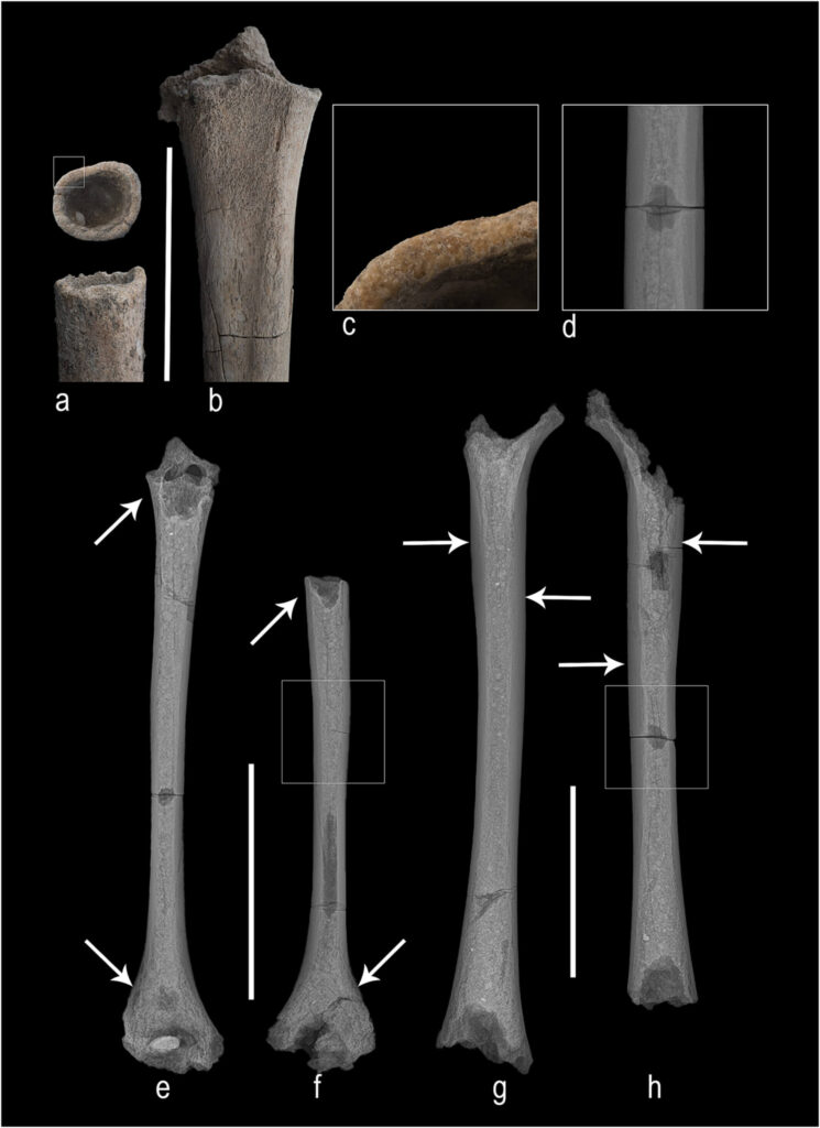
The researchers also considered secondary hyperparathyroidism and thalassemia because several lesions overlap with changes produced by these conditions. Either could help explain the weakened skeleton and increased vulnerability to fractures, but poor preservation prevented a firm diagnosis.
The authors cautiously propose that serious injury or illness may have left the adolescent bedridden or confined indoors for a prolonged period. Reduced exposure to sunlight could then have contributed to vitamin D deficiency. However, this sequence cannot be established from the skeleton, and an existing metabolic condition may instead have made the bones more vulnerable to trauma.
Survival that depended on others
The adolescent’s well-preserved teeth contained no cavities, abscesses or evidence of tooth loss during life. Researchers also found no growth deficit or common skeletal signs of childhood stress, indicating that the young person continued to receive enough nourishment to support broadly normal development.
Chronic jaw dysfunction, fragile bones and recovery from a possible opening in the skull would nevertheless have affected everyday life. The condition may have required specially prepared food, pain relief, restricted physical activity and assistance during periods when the adolescent could not manage essential tasks alone.
The grave lends additional weight to this picture of social inclusion. Its numerous objects, unusually diverse beads and dog burial suggest that the adolescent retained a meaningful place within the community. These offerings cannot reveal a specific status or emotional relationship, but they do not resemble the burial of someone cast aside because of illness.
The researchers acknowledge that poor preservation and concretions covering parts of the skeleton leave room for alternative diagnoses. Trepanation, osteomalacia and medicinal areca use therefore remain interpretations supported to different degrees, not established facts.
Even with those limitations, Burial 3b offers a rare glimpse of illness as a shared experience. The bones record years of injury and physical adaptation; the adolescent’s survival points toward the people who helped make those years possible.
The case may represent evidence of Bronze Age trepanation in Myanmar, but its deeper significance lies elsewhere. It preserves the story of a young person who endured severe physical challenges and a community that appears to have continued caring for them until death.
Cover Image: The skull of Burial 3b shows a circular, healed opening that researchers interpret as possible evidence of trepanation, alongside a separate lesion linked to earlier trauma. Credit: Willis et al. (2026) / International Journal of Osteoarchaeology, CC BY
The open-access study by Anna Willis, Baptiste Pradier, Thu Thu Win and T. O. Pryce was published online on August 17, 2026, in the International Journal of Osteoarchaeology.